| dc.contributor.advisor | Nystad, Mona | |
| dc.contributor.advisor | Villegas, Luis | |
| dc.contributor.author | Nalliah, Maddhusja | |
| dc.date.accessioned | 2021-09-27T08:59:45Z | |
| dc.date.available | 2021-09-27T08:59:45Z | |
| dc.date.issued | 2021-07-01 | en |
| dc.description.abstract | Preeclampsia (PE) is a pregnancy-related disorder affecting 5-8% of women worldwide (4% in Norway). It is believed that placental ischemia is the initial event in the development of PE and is characterized by placental insufficiency and clinical symptoms such as hypertension and proteinuria. In this project, we aimed to study suitable biomarkers for morphological analysis of human term placenta from normal pregnancies and women with PE using advanced fluorescence microscopes.
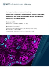
To reach this objective, we optimized the labeling steps for advanced fluorescence optical microscopy imaging of formalin-fixed paraffin-embedded (FFPE) and cryo-preserved tissue sections of the human placenta. Furthermore, morphological and subcellular differences between healthy and preeclamptic placentas were investigated. For this, various fluorescence microscopy techniques were explored, including whole-slide scanner, high-resolution deconvolution microscopy (DV) and super-resolution structured illumination microscopy (SIM), along with diverse image processing tools and analysis of the microscopy images. In this thesis, diverse strategies were examined for the labeling of placental biomarkers including immunofluorescence staining of laeverin, cytokeratin-7 (CK-7) and placental alkaline phosphatase (PLAP), as well as direct labeling of F-actin, membranes and nuclei via phalloidin-Atto 647 N, CellMask Orange (CMO) and DAPI, respectively.
The microscopy investigation revealed actin spots abundantly localized in subtypes of the chorionic villi in both healthy and PE placentas, such as terminal villi (p-value 0.55), mature intermediate villi (p-value 0.50), immature intermediate villi (p-value 0.54) and stem villi (p-value 0.47), thus no observable differences. However, we found significant differences (p-value 0.015) of syncytial knots in PE compared to healthy tissue. A disorganized brushborder at the apical surface seems to be observed in the PE chorionic villi. Moreover, we found PLAP expression in the syncytial microvesicles in healthy placentas. The immunofluorescence study using laeverin and CK-7 antibodies seem to show co-localization in the syncytial plasma membrane in healthy placentas, though the labeled PE tissues showed laeverin expression in the syncytial plasma membrane and cytoplasm, including overexpression of laeverin in the fetal capillaries.
In conclusion, the biomarkers explored in this study may have the potential to play an important role in understanding and predict PE in the future. | en_US |
| dc.identifier.uri | https://hdl.handle.net/10037/22666 | |
| dc.language.iso | eng | en_US |
| dc.publisher | UiT Norges arktiske universitet | no |
| dc.publisher | UiT The Arctic University of Norway | en |
| dc.rights.holder | Copyright 2021 The Author(s) | |
| dc.rights.uri | https://creativecommons.org/licenses/by-nc-sa/4.0 | en_US |
| dc.rights | Attribution-NonCommercial-ShareAlike 4.0 International (CC BY-NC-SA 4.0) | en_US |
| dc.subject.courseID | MBI-3911 | |
| dc.subject | VDP::Medisinske Fag: 700::Klinisk medisinske fag: 750 | en_US |
| dc.subject | VDP::Medical disciplines: 700::Clinical medical disciplines: 750 | en_US |
| dc.subject | Human term placenta | en_US |
| dc.subject | Preeclampsia | en_US |
| dc.subject | Advanced fluorescence optical microscopy | en_US |
| dc.subject | Super-resolution | en_US |
| dc.subject | Morphology study | en_US |
| dc.subject | Biomarkers | en_US |
| dc.subject | VDP::Medisinske Fag: 700::Basale medisinske, odontologiske og veterinærmedisinske fag: 710::Medisinsk molekylærbiologi: 711 | en_US |
| dc.subject | VDP::Medical disciplines: 700::Basic medical, dental and veterinary science disciplines: 710::Medical molecular biology: 711 | en_US |
| dc.subject | VDP::Teknologi: 500::Medisinsk teknologi: 620 | en_US |
| dc.subject | VDP::Technology: 500::Medical technology: 620 | en_US |
| dc.subject | Morkake | en_US |
| dc.subject | Svangerskapsforgiftning | en_US |
| dc.title | Optimization of biomarkers for morphological analysis of healthy and preeclamptic term human placental tissue sections using advanced fluorescence microscopy methods | en_US |
| dc.type | Mastergradsoppgave | no |
| dc.type | Master thesis | en |


 English
 norsk
norsk


